Theme: Exploring New Advancements in Ophthalmology
Euro-Ophthalmology 2024
- Welcome Message
- Conference Sessions
- Conference Relevant Keytopics
- Market Analysis
- Related conference & Societies
We are elated to invite you to the 38th European Ophthalmology Congress which is going to be held in every one’s dream destination London, UK.
Join many of the ophthalmologists, optometrists, eye surgeons, eye care specialists, physicians, radiologists, medical imaging specialists, clinical researchers & scientists, professors, ophthalmology equipment companies, medical practitioners, diagnostic instruments in optometry and all the professionals from various multispecialty areas as they come together to share, partake research ideas, explore new collaborative, build career beginnings and witness the finest of the ophthalmology research community has to offer.
Why do attendees come to Ophthalmology Conferences year after year through their professions? There are so many reasons. Here are the top few:
- Get the latest updates in technology and management of ophthalmology.
- Direct interaction with our industry colleagues from around the world.
- Play a role in mentoring the afterward generation of physician-scientists and academic leaders.
- Bring home the latest up-to-dates on emerging technologies, clinical applications and practical solutions.
- Estimate which products will be best for your organization as lots of exhibitors showcase their products and services.
Track 01: Ophthalmology
Ophthalmology is a surgical subspecialty within medicine that deals with the diagnosis and treatment of eye disorders and it deal both primary and specialty eye care that encompasses many different subspecialties, including: strabismus/pediatric ophthalmology, glaucoma, neuro-ophthalmology, retina/uveitis, anterior segment/cornea, oculoplastic/orbit, and ocular here we mentioned few of them.
Track 02: Glaucoma: A Vision Loss
Glaucoma is a chronic, progressive, degenerative disorder of the optic nerve that produces characteristic visual field damage. Glaucoma is the second cause of blindness, and importantly it is irreversible. Any group of related eye disorders characterized by increased pressure within the eye which impairs the vision and may slowly cause eye damage and total loss of vision. In many cases glaucoma may be asymptomatic, meaning it shows no symptoms half of those living with glaucoma are unware that they are affected.
Track 03: Retina and Retina Detachement
The retina is the layer of specialized nerve tissue lining the back of the eye that allows you to see. The retina is very loosely attached to the back of the eye and in some cases, it may become detached. When the retina detaches, it is lifted from its normal position by fluid that accumulates between the retina and the eye wall. A detached retina is disconnected from its oxygen and nutrition supply. A hole or tear in the retina causes a retinal detachment. The presence of this hole or tear allows fluid to get underneath the retina and displace it from its nutrition source. Retinal detachments can develop at any age but tend to occur more commonly in the elderly. Retinal detachments usually occur around the time that the vitreous separates from the retina (PVD). Most PVDs causes no long-term damage to the eye, but in some individuals, they result in retinal tears that can then lead to a retinal detachment.
Track 04: Cornea Disorders and Treatments
The cornea is the outermost clear, transparent integral layer located in the front of the iris and pupil. It is responsible for the involuntary eyelids reflex and helps to focus the light on the retina. Any change in the cornea layer consistency causes altered vision. With a decrease in transparency of this layer, blurred vision can occur.
Treatments might include medications, laser treatment, or surgery, depending on the condition. Infections are treated with medicated eyedrops (antibiotics, antivirals, and antiparasitics) and, in some cases, oral medication. Herpetic stromal keratitis is a recurring swelling that develops after a herpes eye infection and is managed with anti-inflammatory steroid eye drops.
An abrasion might require temporary patching or a bandage contact lens, depending on the cause and extent of the injury.
Track 05: Ocular Oncology
Ocular oncology is the branch of medicine dealing with tumours relating to the eye and its adnexa. Eye cancer can affect all parts of the eye. Eye neoplasms are benign or malignant (cancerous) tumours that occur in or around the eye. Eye cancer is rare in comparison to other cancerous tumours. Benign tumours may also be called dermoid cysts. Malignant tumours may be either rhabdomyosarcoma or retinoblastoma, and can be primary, starting within the eye, or secondary (metastatic), spreading to the eye from another organ. Most eye tumours are metastatic— usually starting in the breast, lung, bowel or prostate.
Two types of primary tumours develop within the eye itself
- Retinoblastoma in children
- Ocular melanoma in adults.
Retinoblastoma: Retinoblastoma is a cancer of the eye that begins in the retina, the eye’s light-sensitive tissue. It is a rare form of cancer, though the most common eye cancer that affects children under five years of age.
Ocular Melanoma: Ocular melanoma is a rare form of cancer that occurs in the pigment producing cells of your eyes. Ocular melanomas usually develop in the middle of the three layers of your eye, called the uvea. In rare cases, the melanoma can also develop on the conjunctiva. When tumours develop inside and around the eye, a specialized team of doctors— including ophthalmologists, oncologists, radiation specialists, and surgeons are called upon to provide the best possible care for preservation of life, vision, and cosmetic appearance.
Track 06: Pediatric Ophthalmology
Pediatric ophthalmology is a branch of ophthalmology that focuses on treating children’s vision problems and eye illnesses. Childhood is a crucial time for proper eye care because many visual issues arise as the eyes develop and could impact the child’s future development.
Track 07: Dry Eye & Keratoconus
Dry eye disease (also known as dry eye syndrome) is a common condition that can affect your quality of life. Dry eyes can make your eyes feel sore and gritty and make your vision blurry. If you have dry eye disease, your eyes my feel sensitive to light, they may sting or burn, or look red. Your eyelids may be sticky when you wake up. Sometimes dry eye disease can cause excess tearing. You might also have blepharitis (inflamed eyelids). You may feel a sensation of something in your eye and you may have difficulty wearing contact lenses. Your tear film is made up of 3 layers: fatty oils, watery tears and mucus. When your tears are working normally these 3 components keep your eyes smoothly lubricated. If you have a problem with 1 of these 3 layers you may develop dry eye disease.
Normally your cornea, the clear outer lens or "windshield" of the eye, has a dome shape, like a ball. Sometimes the structure isn’t strong enough to hold its round shape and it bulges outward, like a cone. This is called keratoconus.
It impossible for your eye to focus without glasses or contact lenses. In fact, you may need a corneal transplant to restore your sight if the condition gets bad enough.
A treatment called cornea collagen crosslinking may stop the condition from getting worse. Or your doctor could implant a ring called an Intacs under the cornea’s surface to flatten the cone shape and improve vision.
Track 08: Neuro-Ophthalmology
Neuro-ophthalmology is an ophthalmic subspecialty that merges the fields of ophthalmology and neurology. This super speciality field deals with neurological problems associated with the eyes and also deal with the nervous system’s many complex diseases that affect vision, eye alignment, and many optic nerve disorders. They have a unique ability to diagnose and evaluate patients from medical, ophthalmic, and neurologic standpoints to treat neuro-ophthalmologic disorders.
Track 09: Ophthalmology Surgery
Ophthalmology surgery is also known as ocular surgery. Ophthalmologic surgery is a surgical procedure performed on the eye or any part of the eye. Surgery on the eye is routinely performed to repair retinal defects, remove cataracts or cancer, or to repair eye muscles. The most common purpose of ophthalmologic surgery is to restore or improve vision.
Track 10: Diabetic Retinopathy
Diabetic Retinopathy (DR) is the leading cause of vision deterioration and blindness across the globe. It is a complication of diabetes affecting the eyes. Among the almost 300 million people who have diabetes, about one-third of them develop Diabetic Retinopathy (DR). Read on to find out what diabetic retinopathy is, its risks and symptoms, whether it is reversible, and how to live with this disease.
Track 11: Refractive Surgery
Refractive surgery can correct refractive errors like Myopia, Hyperopia and astigmatism. Some of these surgeries reshape the cornea. Others implant a lens in your eye. These surgeries focus light correctly on the retina so you can see more clearly.
The most well-known refractive mistakes in youngsters are:
- Myopia (otherwise called near-sightedness)
- Hyperopia (additionally called farsightedness)
- Astigmatism (contorted vision)
It is conceivable to have at least two kinds of refractive mistake in the meantime.
Near-sightedness: A near-sighted eye is longer than ordinary or has a cornea that is excessively steep, with the goal that the light beams center before the retina. Close protests look clear, however removed items seem obscured.
Hyperopia: A hyperopic eye is shorter than typical. Light from close protests can't center obviously around the retina. The words on a page will appear to be hazy, or it will be hard to see all around ok to do quit for the day, such as threading a needle.
Astigmatism: Astigmatism contorts or obscures vision for both close and far items. It's relatively similar to investigating a fun house reflects in which you show up excessively tall, too wide or too thin. A typical cornea is round and smooth, similar to a ball. It is conceivable to have astigmatism in blend with near-sightedness or hyperopia.
Track 12: Strabismus
Strabismus is a failure of the two eyes to maintain proper alignment and work together as a team. One eye turns inwards, upwards, downwards, or outwards, while the other one focuses at one spot. This typically happens as a result of the muscles that management the movement of the attention and therefore the protective folds, the extra ocular muscles, aren’t operating along.
Track 13: Uveitis
Uveitis could be a form of inflammation to the center layer of the attention (uvea), structure typically contains the iris, membrane and choroid coat. The causes of uveitis area unit of various varieties starting from a straightforward microorganism Infection to severe eye injury, studies show that the presence of inflammatory disease, AIDS, skin condition will increase the probabilities of uveitis. During this the inner a part of the attention turns to red in color, thanks to this pain, blurred vision and picture sensitivity happens. Prompt use of anti-bacterial will treat the condition otherwise their area unit high probabilities that it should cause eye disease, cataract etc..,
Track 14: Optical Technologies and Laser Science in Ophthalmology
Light, like radio, consists of electromagnetic waves. The major difference between the two is that light waves are much shorter than radio waves. The use of electromagnetic waves for long-distance communications was the beginning of an industry known first as wireless and later as radio. This industry was the foundation for electronics, which brought the world so many fascinating technologies. The word LASER stands for Light Amplification by Stimulated Emission of Radiation. A laser is a device that emits a concentrated beam of photons, which are the basic units of electromagnetic radiation.
Track 15: Conjunctivitis
Conjunctivitis, or pink eye, is an irritation or inflammation of the conjunctiva, which covers the white part of the eyeball. It can be caused by allergies or a bacterial or viral infection. Conjunctivitis can be extremely contagious and is spread by contact with eye secretions from someone who is infected.
Symptoms include redness, itching and tearing of the eyes. It can also lead to discharge or crusting around the eyes. It's important to stop wearing contact lenses whilst affected by conjunctivitis. It often resolves on its own, but treatment can speed the recovery process. Allergic conjunctivitis can be treated with antihistamines. Bacterial conjunctivitis can be treated with antibiotic eye drops.
- By skin-to-skin contact
- By touching a contaminated surface
Track 16: Cataract
A cataract is a clouding of the natural lens of the eye. This clouding of the eye prevents light rays from entering the lens and focusing on the retina. The retina is a light-sensitive tissue lining of the eye and is located at the back of the eye. This cloudiness occurs when some of the proteins from the eye's lens start to alter their shape and obstruct the vision.
There are three basic techniques for cataract surgery
1. Phacoemulsification: This is the most common form of cataract removal as explained above. In this most modern method, cataract surgery can usually be performed in less than 30 minutes and usually requires only minimal sedation and numbing drops, no stitches to close the wound, and no eye patch after surgery.
2. Extracapsular cataract surgery: This procedure is used mainly for very advanced cataracts where the lens is too dense to dissolve into fragments (phacoemulsify) or in facilities that do not have phacoemulsification technology. This technique requires a larger incision so that the cataract can be removed in one piece without being fragmented inside the eye. An artificial lens is placed in the same capsular bag as with the phacoemulsification technique. This surgical technique requires a various number of sutures to close the larger wound, and visual recovery is often slower. Extracapsular cataract extraction usually requires an injection of numbing medication around the eye and an eye patch after surgery.
3. Intracapsular cataract surgery: This surgical technique requires an even larger wound than extracapsular surgery, and the surgeon removes the entire lens and the surrounding capsule together. This technique requires the intraocular lens to be placed in a different location, in front of the iris. This method is rarely used today but can be still be useful in cases of significant trauma.
Track 17: Macular Degeneration
Macular degeneration is an eye disease that affects central vision. This means that people with macular degeneration can’t see things directly in front of them. This common age-related eye condition mostly occurs in people over the age of 50.
Macular degeneration affects your macula, the central part of your retina. Your retina is in the back of your eye and controls central vision. People with macular degeneration aren’t completely blind. Their peripheral vision (ability to see things off to the sides) is fine.
Track 18: Latest equipment’s used for ophthalmology surgeries
Ophthalmology is a branch of medicine deals with such significant diseases as cataracts, blindness, glaucoma and macular degeneration. In order for ophthalmologists to provide the patients with effective and responsible treatment, it is crucial to use ophthalmology equipment made in accordance to high quality standards.
Technological advancements are playing a pivotal role in improving the early detection and management of glaucoma: Advanced Imaging Techniques: Optical coherence tomography (OCT) has transformed the assessment of optic nerve and retinal nerve fibre layer thickness, aiding in the early diagnosis of glaucoma.
Ophthalmology Conference | Eye Conferences | Retina Conferences | glaucoma Society | European Ophthalmology Congress | Ophthalmology Events | Vision Science Conferences | Pediatric Ophthalmology Conferences | Optometry Conferences |
Top Ophthalmology Conferences | World Ophthalmology Congress | Best Ophthalmology Conferences | International Ophthalmology Summit | Upcoming Ophthalmology Meetings | Ophthalmology Events
The global ophthalmology market is expected to gain market growth in the forecast period of 2023 to 2030. Data Bridge Market Research analyses that the market is growing with a CAGR of 6.4% in the forecast period of 2024 to 2030 and is expected to reach USD 84,655.95 million by 2030, from USD 51,459.32 million in 2022
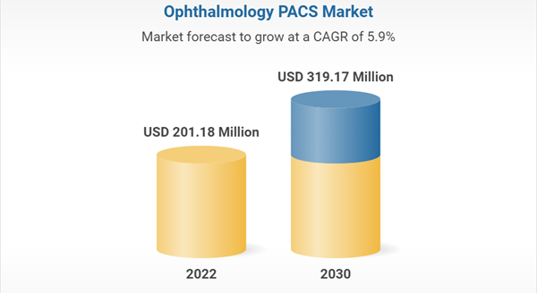
Ophthalmology is a branch of medicine that deals with the anatomy, physiology, treatment and surgery of the visual pathways of the eye, including surrounding areas and the visual aspects of the brain. Ophthalmic devices are medical equipment designed for surgery, diagnosis, and vision correction.
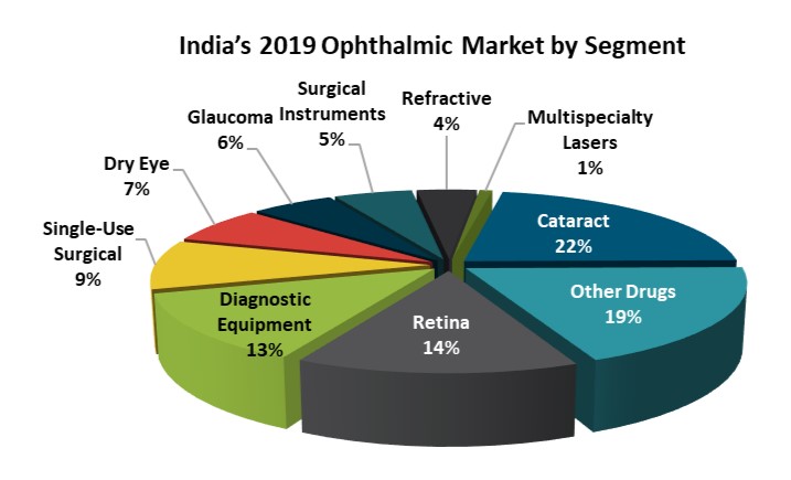
The devices gained increased importance and adoption due to the high prevalence of several ophthalmic diseases, such as glaucoma, cataract, and other vision-related issues. Ophthalmic drugs are medications that are specially designed and administered for the treatment of various eye-related disorders, including colour blindness, diabetic macular edema, cytomegalovirus (CMV), and age-related macular degeneration (AMD).
Related Conferences: Ophthalmology Conferences | Vision Conferences | Eye Conferences 2023 | Global Ophthalmology Meetings | Optometry Conferences | Glaucoma conferences | Ophthalmologists Conferences | Clinical Ophthalmology Conferences | Vision Science Conferences | Vision Congress
9th International Conference on Eye and Vision February 22-23, 2024 Zurich, Switzerland; 5th World congress on Ophthalmology and Optometry March 14-15, 2024 London, UK; 38th European Ophthalmology Congress March 14-15, 2024 London, UK; 5th World Congress on Ophthalmology and Vision Science March 18-19, 2024 Zurich, Switzerland; 22nd Global Ophthalmologists Annual Meeting April 25-26, 2024 London, UK
Related Societies:
USA: American Society of Retina Specialists, Retinal American Academy of Ophthalmology, Optometric Retina Society, Pan America Glaucoma Society, American Glaucoma Society
Europe: European Society of retina Specialists, European Vitroretinal Society, European Glaucoma Society, European Society of cataract and refractive surgeons, European Mucosal Immunology Group
Asia Pacific: Japanese society of Cataract and refractive Surgeons, Asia Pacific Vitro retinal Societies, Asia Cornea Society, Afro-Asian Council of Ophthalmology , Asian Angle-Closure Glaucoma Club
Conference Highlights
- Ophthalmology
- Glaucoma: A Vision Loss
- Retina and Retinal Detachment
- Cornea Disorders and Treatments
- Ocular Oncology
- Pediatric Ophthalmology
- Dry Eye & Keratoconus
- Neuro-Ophthalmology
- Ophthalmology Surgery
- Diabetic Retinopathy
- Refractive Surgery
- Strabismus
- Uveitis
- Optical Technologies and Laser Science in Ophthalmology
- Conjunctivitis
- Cataract
- Macular Degeneration
- Latest equipments used for ophthalmology surgeries
To share your views and research, please click here to register for the Conference.
To Collaborate Scientific Professionals around the World
| Conference Date | March 14-15, 2024 | ||
| Sponsors & Exhibitors |
|
||
| Speaker Opportunity Closed | |||
| Poster Opportunity Closed | Click Here to View | ||
Useful Links
Special Issues
All accepted abstracts will be published in respective Our International Journals.
- Journal of Clinical & Experimental Ophthalmology
- International Journal of Ophthalmic Pathology
- Journal of Lasers, Optics & Photonics
Abstracts will be provided with Digital Object Identifier by


